Nisha Pal
Research Scholar, Suresh Gyan Vihar University, Jaipur, India
Abstract: Aim of presenting this paper is to detect the brain tumor by using thresholding and watershed algorithm. In order to find the correct size, shape, limit extraction and area of tumor Brain tumor identification can be helpful. To detect and segment brain tumor there are three stages. For detection of tumor based on segmentation there is an efficient algorithm. For detection of tumor in scanned image, firstly, enhancement process of scanned image and morphological operators are applied. Once edges are detected , operators are applied so that the boundaries can be extracted and the size of tumors can be found.
Keywords — Brain Tumor, MRI, Segmentation.
- INTRODUCTION
The brain is a delicate, sensitive, irreplaceable and lightweight tissue. It is a steady place for examples to enter and settle among one another. A tumor is essentially a swelling of a part of the body that becomes a wild force that governs the typical forces of its development. Brain tumor is a gathering of irregular cells that becomes either inside the mind or on the other hand around the brain. All healthy brain cells could be destroy by tumors. It can indirectly, adversely harm the healthy brain cells by swarming into different parts of the brain and causing aggravation, brain swelling and inducing pressure inside the skull. [1]
Exact measurements in brain tumor diagnosis are very troublesome due to assorted shapes, sizes and tumor appearance. Tumors may develop unexpectedly, leading to adjacent neighboring. In addition, the organization also provides a large number of unusual structures for solid tissue. In this paper, we will use a division related to morphological activities to establish a three-dimensional segmentation process for brain tumors. [2]
Types of brain tumor:-
Tumor
Tumor is also termed as ‘Neoplasm’. Tumors are completely unexpected cancers.
Tumors are of three types:-
1) Benign (Non- Cancerous)
2) Pre-Malignant
3) Malignant (Cancerous)
Benign Tumor
Benign tumor is a mass of cells , that forms a lump, which lacks the ability to invade the neighboring tissues or spread to other non-neighboring parts of the body. These tumor have a much slower growth rate and therefore, they are much less risky. Benign brain tumors can usually be successfully removed with surgery and don’t usually grow back.
Warts are a normal condition of benign tumors.
Pre-Malignant Tumor
Premalignant Tumor is the precancerous stage of tumor and is considered a disease that can cause cancer if not treated properly.
Malignant Tumor
Malignant is the type of tumor that grows over time and eventually the result of the tumor is human death. A malignant disease is essentially a medical term describing a serious disease. Malignant tumors are terms that are often used to describe tumors. [2]
Magnetic Resonance Imaging (MRI)
Magnetic resonances imaging (MRI), [also known as, Magnetic Resonance Tomography (MRT), Nuclear MRI (NMRI)] is a medical imaging method used in radiology to look closely at images of the internal structure of the body. [2]
MRI utilizes three electromagnetic fields: the electrostatic field, which is an extremely robust static attraction field that energizes the hydrogen nucleus; the gradient field is a more fragile time-shifting field for spatial coding; and is used to control the hydrogen nucleus. A weak RF field that provides a measurable signal is collected through the Radio Frequency antenna. [3]
This procedure is mainly used to distinguish between the differences in the tissues which have a evidently better strategy compared to computer tomography. Hence, this makes this an extraordinary procedure for the identification of brain tumor and cancer imaging. [1]
- RELATED WORK
Watershed and threshold techniques contribute to the division of brain tumors. The segmentation of an image depends on the image being segmented into regions.
Image segmentation is based on comparison quality. Those with Similarities are placed out into groups, so that we could get the features and important information regarding the image.
Threshold Segmentation:
Threshold segmentation is the based on a simple segmentation technique where we input image called the gray scale image is first converted into a binary format.
This segmentation method is carried out on a threshold value which varies as per the features of the gray scale image while being converted into a binary image format. The selection of a threshold value for segmentation, is the prime concern. Image utilizing histogram helps us in finding a single threshold value for the same. Image Utilizing Histogram is a sort of histogram that is based as a graphical representation for the tonal distribution in a advance picture. The quantity of pixels for each tonal value is plotted on the histogram.
Watershed segmentation:-
Watershed segmentation is extraordinary, compared to other strategies for collecting image pixels based on their intensity. Pixels at the same intensity are combined, which eases the segmentation procedure so that we can isolate a tumor from the image. Watershed is a scientific morphological working tool.
Detection of Brain Tumor
After having performed the segmentation procedure by any of the above mentioned method , be it watershed or the threshold , we are left with the most awaited part on which we need to work and that is the tumor .This bit contains just high exceptional pixels which appears thoroughly its white part.
Extraction of Boundry in Brain Tumor
Edge-based segmentation is the most widely accepted technique, which depends on the discovery of edges, ie the separation of different regions.
Shape and Size of Brain Tumor
Once we have the boundary of the tumor, shape of the tumor can easily be decided. If we getting round boundry then its shape is roundabout and so on.
After that we are finding the size of the brain tumor, it is Estimated in the matrix form (m*n). [2]
III. EXPERIMENTAL RESULT
For this we will use image of magnetic resonance images (MRI) by using processing of the image.
Input Image:
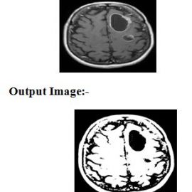
After all operation we detect the tumor area.
- CONCLUSION
In this paper, description of Brain tumor detection and segmentation by using Watershed and thresholding algorithm has been put down.
The test results appeared in the upper region, and the canny edge detection operator showed the accuracy of the tumor and found the boundary of the tumor. The shape and size of the tumor are depicted.
REFERENCES
[1].Rachana Rana, H.S. Bhadauria, Annapurna Singh3“International Journal of Emerging Technology and Advanced Engineering”. ISSN 2250-2459, ISO 9001:2008 Certified Journal, Volume 3, Issue 6, June 2013
[2]. Roshan G. Selkar, Prof. M. N. Thakare, “BRAIN TUMOR DETECTION AND SEGMENTATION BY USING THRESHOLDING AND WATERSHED ALGORITHM, ISSN 2348 – 9928, IJAICT Volume 1, Issue 3, July 2014.
[3]. Rohini Paul Joseph1, C. Senthil Singh2, M.Manikandan3 “BRAIN TUMOR MRI IMAGE SEGMENTATION AND DETECTION IN IMAGE PROCESSING”. IJRET: International Journal of Research in Engineering and Technology eISSN: 2319-1163 | pISSN: 2321-7308